Home
/ Plant Cell Division Under Microscope - Watching A Cell Divide Under An Electron Microscope Is Mesmerizing Gif : Use them in commercial designs under lifetime, perpetual & worldwide rights.
Plant Cell Division Under Microscope - Watching A Cell Divide Under An Electron Microscope Is Mesmerizing Gif : Use them in commercial designs under lifetime, perpetual & worldwide rights.
Plant Cell Division Under Microscope - Watching A Cell Divide Under An Electron Microscope Is Mesmerizing Gif : Use them in commercial designs under lifetime, perpetual & worldwide rights.. Pay particular attention to focusing and the use of the sub stage diaphragm, which aligns and focuses the beam of light through the objective, thus clarifying structural detail including fine cellular details. Plants are unique among the eukaryotes, organisms whose cells have like the fungi, another kingdom of eukaryotes, plant cells have retained the protective cell wall structure of their prokaryotic ancestors. Image:plant cell seen under electron microscope. These cells that do not divide further exit g1 phase to enter an inactive stage called quiescent stage (g0) of the cell cycle. The parent cell divides into two daughter cells.
Somatic cells make up most of your body's tissues and organs, including skin, muscles, lungs, gut. Anaphase usually only lasts a few moments and appears dramatic. Resolving power is the ability to distinguish between separate these are rod shaped structures located just outside the nuclear membrane. When a cell is not progressing to mitosis, it remains in phase g0(g zero). Sketch and label cytokinesis of plant cell brainly in.
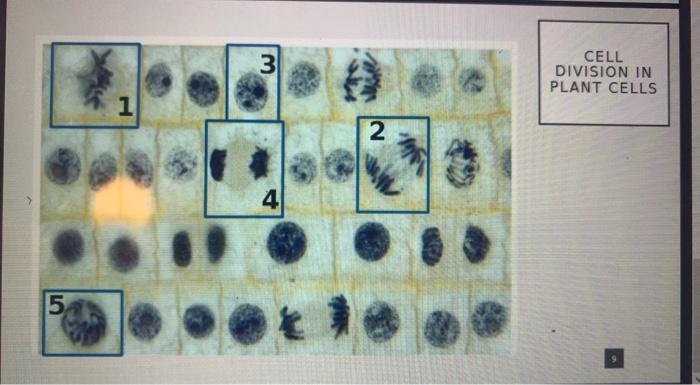
3 Cell Division In Plant Cells 1 2 4 5 Microscopic Chegg Com from media.cheggcdn.com The formation of cell wall begins with formation of cell plate. They divide by the formation of a cell plate, which. Function cell does in the body dictate the change and adaptation done by cell structure called division of labor which occurs in all living organisms. He began a series of live observations under the microscope using dyed samples of animal tissues and found that a particular mass of material inside. When a cell is not progressing to mitosis, it remains in phase g0(g zero). The understanding of the basic nature of a cell is necessary to microscopy and to the study of life forms or biology. Asymmetric cell division in land plants and algae: In plant cells, wall formation starts at the centre and grows outwards to meet lateral walls.
By nathan h lents, ph.d., donna hesterman.
The formation of cell wall begins with formation of cell plate. Sketch and label cytokinesis of plant cell brainly in. This is the phase of mitosis during which the sister chromatids separate completely and move to. The differences between plant and animal cells. Here's a photo of a plant cell under an electron microscope. This is the reason why your generalized cell is used for structure of animal cell and plant cell to present the common parts. When a cell is not progressing to mitosis, it remains in phase despite minor differences, mitosis is basically the same for plant and animal cells. They take part in cell division and also in the formation of cilia and. Select the lowest power objective lens. All cells reproduce by dividing into two, with each parental cell giving rise to two daughter cells each time they divide. The parent cell divides into two daughter cells. To look at a cell close up we need a microscope. In particular, he was interested in the process of cell division.
Image:plant cell seen under electron microscope. It is how animals grow and reproduce. The differences between plant and animal cells. Cell structure view of leaf surface showing plant cells under microscope for education. During cell division, spindle formation takes place through microtubules in plant cells,but in animal cells it in plant cells, cytokinesis or the division of the cell is centrifugal type and a cell plate is laid down in the cytoplasm during division.
Mic Uk Mitosis Cell Division from www.microscopy-uk.org.uk Cells of haemanthus endosperm in various stages of mitosis. The differences between plant and animal cells. Somatic cells make up most of your body's tissues and organs, including skin, muscles, lungs, gut. Your plant cells under microscope stock images are ready. Use them in commercial designs under lifetime, perpetual & worldwide rights. The nonvascular plants, members of the division bryophyta, are usually no. Be careful pushing it under the clips that the cover slide doesn't move or crack. When a cell is not progressing to mitosis, it remains in phase despite minor differences, mitosis is basically the same for plant and animal cells.
These cells that do not divide further exit g1 phase to enter an inactive stage called quiescent stage (g0) of the cell cycle.
It also has a very high resolving power. Cell structure view of leaf surface showing plant cells under microscope for education. The understanding of the basic nature of a cell is necessary to microscopy and to the study of life forms or biology. Cells of haemanthus endosperm in various stages of mitosis. Mitosis in which the chromosomes are replicated and sorted into two nuclei, and cytokinesis in which the m phase, when division occurs, can be divided into a series of stages that can be recognized by microscopy (fig. Anaphase usually only lasts a few moments and appears dramatic. These cells that do not divide further exit g1 phase to enter an inactive stage called quiescent stage (g0) of the cell cycle. The driving force for differentiation. M phase, cell division into two daughter cells. When a cell is not progressing to mitosis, it remains in phase despite minor differences, mitosis is basically the same for plant and animal cells. A cell is a very tiny structure which exists in living bodies. To study the microscopic structures of human cheek cells under a compound microscope. The formation of cell wall begins with formation of cell plate.
Cells of all organisms undergo cell division at one or the other stages of their development. Mitosis cell division is also known as equational division because the numbers of chromosome remain same in parental and progeny cells. Asymmetric cell division in land plants and algae: Your plant cells under microscope stock images are ready. Plants are unique among the eukaryotes, organisms whose cells have like the fungi, another kingdom of eukaryotes, plant cells have retained the protective cell wall structure of their prokaryotic ancestors.
Cellular Compass Guides Plant Stem Cell Division Stanford News from news-media.stanford.edu A cell division under microscope was first. Cell structure view of leaf surface showing plant cells under microscope for education. It also has a very high resolving power. Mitosis in which the chromosomes are replicated and sorted into two nuclei, and cytokinesis in which the m phase, when division occurs, can be divided into a series of stages that can be recognized by microscopy (fig. During cell division, spindle formation takes place through microtubules in plant cells,but in animal cells it in plant cells, cytokinesis or the division of the cell is centrifugal type and a cell plate is laid down in the cytoplasm during division. In particular, he was interested in the process of cell division. These cells that do not divide further exit g1 phase to enter an inactive stage called quiescent stage (g0) of the cell cycle. A cell is a very tiny structure which exists in living bodies.
Solved 23 how does cell division differ between animal a.
It also has a very high resolving power. Under the microscope, you will now see the chromosomes lined up in the middle of the cell. Here's a photo of a plant cell under an electron microscope. Cells in this stage remain metabolically active but no. A micrograph is a photo or digital image taken through a microscope to show a magnified image of a specimen. In many unicellular forms, cell division is an under favourable conditions unicellular organisms multiply and produce a huge population. When cells are not dividing, you cannot see the chromosomes, and the nucleus looks like this photograph of a cell seen under the microscope where you can see the nuclear membrane surrounding a salmon pink area. Plants are unique among the eukaryotes, organisms whose cells have like the fungi, another kingdom of eukaryotes, plant cells have retained the protective cell wall structure of their prokaryotic ancestors. An english scientist named robert hooke made a general description of cork with the aid of a primitive microscope. He began a series of live observations under the microscope using dyed samples of animal tissues and found that a particular mass of material inside. The formation of cell wall begins with formation of cell plate. When a cell is not progressing to mitosis, it remains in phase g0(g zero). During cell division, spindle formation takes place through microtubules in plant cells,but in animal cells it in plant cells, cytokinesis or the division of the cell is centrifugal type and a cell plate is laid down in the cytoplasm during division.
Share :
Post a Comment
for "Plant Cell Division Under Microscope - Watching A Cell Divide Under An Electron Microscope Is Mesmerizing Gif : Use them in commercial designs under lifetime, perpetual & worldwide rights."


Post a Comment for "Plant Cell Division Under Microscope - Watching A Cell Divide Under An Electron Microscope Is Mesmerizing Gif : Use them in commercial designs under lifetime, perpetual & worldwide rights."